The thrombin (IIa) catalyzed cleavage of soluble fibrinogen (Fbg) to form fibrin (Fbn) is the terminal proteolytic event in the coagulation cascade. These soluble Fbn monomers spontaneously polymerize to form an insoluble Fbn network which is stabilized by the factor XIIIa catalyzed crosslinking of lys and glu residues of α and γ chains. This Fbn network is the major protein component of the hemostatic plug.
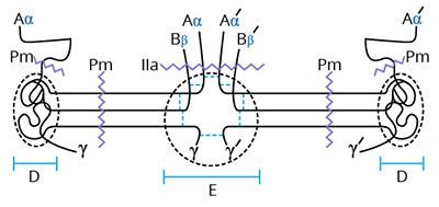
Plasma fibrinogen is large glycoprotein (Mr=340,000) synthesized in the liver and circulating at a concentration of 2.6 mg/ml. It is a disulfide linked dimer composed of 3 pairs of disulfide linked non-identical polypeptide chains (Aα, Bβ and γ). Notable features of the Aα chain are the N-terminal peptide (fibrinopeptide A (FPA, 1-16)), factor XIIIa crosslinking sites and 2 phosphorylation sites. When synthesized, Fbg is fully phosphorylated, but circulates at only 20-30% phosphorylation. The Bβ chain contains fibrinopeptide B (FPB, 1-14), one of the 3 N-linked carbohydrate moieties (Mr=2500) and an N-terminal pyroglutamic acid. The γ chain contains the other N-linked glycosylation site and a factor XIIIa crosslinking sites. The 2 elongated subunits ((AαBβγ)2) are aligned in an antiparallel manner forming a trinodular arrangement of the six chains. The nodes are formed by disulfide rings between the 3 parallel chains. The central node (n-disulfide knot, E domain) is formed by the N-termini of all six chains held together by 11 disulfide bonds. This region contains the 2 IIa-sensitive sites. The release of FPA by cleavage at R16-G17 generates Fbn I, exposing a polymerization site (17-20) on the Aα chain. These regions bind to complimentary regions on the D domain of Fbn to form protofibrils. Subsequent IIa cleavage of FPB (R14-G15) from the Bβ chain exposes additional polymerization sites and promotes lateral growth of the Fbn network.
Each of the 2 domains between the central node (E domain) and the C-terminal nodes (D domain) is composed of parallel α-helical regions of the Aα, Bβ and γ chains coiled around each other to form a “coiled coil” with polar residues directed outward and nonpolar residues forming a hydrophobic core. In this region, all 3 chains possess a protease (plasmin) sensitive site. The other major plasmin sensitive site is in the hydrophilic preturbance of the α-chain from the C-terminal node. Controlled plasmin degradation at these sites converts Fbg into fragment D and fragment E. The individual fragments are isolated by salt fractionation, gel filtration and ion exchange chromatography. The fragments are supplied lyophilized for storage at 4°C. Highly purified research grade fibrinogen (>95% clottable) is prepared by a combination of conventional and affinity techniques. It is supplied as a frozen solution in ctirate-phosphate for storage at -80°C.