Domain Structure of Thrombospondin
The domain structure of the thrombospondin monomer is represented. The thrombospondin monomer is a 150,000 molecular weight glycopeptide. Regions containing amino acid sequence homologous to procollagen, properdin and epidermal growth factor (EGF) are shown. Heparin, calcium ion, and cell binding domains are indicated. Native thrombospondin is a 450,000 molecular weight homotrimer consisting of three identical peptide chains linked through interchain disulfide bonds.

Showing the single result

  • Human Thrombospondin (TSP-1)

    HCTP-0200

    $577.00$5,070.00

    SKU: HCTP-0200 Category:
    Price:$5,070.00/1 mg, $577.00/100 µg
    Size 1 mg, 100 µg
    Formulation 50% glycerol/water (v/v)
    Storage -20°C
    Shelf Life 12 months
    Purity >95% by SDS-PAGE
    Activity Determination N/A

Thrombospondin is a high-molecular weight, heparin-binding glycoprotein constituent of human platelets (1). Ranging from 30-50 µg per 109 platelets, thrombospondin is one of the most abundant proteins in the platelet a-granule (2,3). Thrombospondin was initially termed “thrombin-sensitive protein” based upon its release by thrombin-activated platelets (4,5). Structurally, thrombospondin is a 450,000 molecular weight glycoprotein (1) consisting of three, identical, disulfide-linked polypeptide chains (6-8). The binding of thrombospondin to the surface of both resting and thrombin-activated platelets has been reported (9). A thrombospondin-specific membrane receptor has also been partially characterized (10). Functionally, platelet-derived thrombospondin may play a role in platelet adherence and aggregation (11).

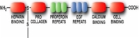
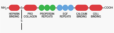
Thrombospondin is not an exclusive product of platelets and megakaryoctes. The synthesis of thrombospondin by endothelial cells (12), fibroblasts (13), monocytes and macrophages (14), and osteoblasts (15) has been reported. Thrombospondin is also an integral component of the basement membrane in a number of different tissues (16). Thrombospondin interacts with a variety of extracellular macromolecules including heparin (1,17), collagen (18), fibrinogen and fibronectin (19), plasminogen (20), plasminogen activator (21), and osteonectin (22). Through the collective efforts of a number of different investigators employing both peptide chemistry and cDNA analytical techniques, distinct heparin, Ca2+-ion, platelet, and protein binding domains within thrombospondin have been identified (23). Based upon its specific interactions with both cells and extracellular matrix components, thrombospondin has been hypothesized to be a member of a class of extracellular proteins which may modulate cell-matrix interactions (24).

Our human thrombospondin (TSP-1) is prepared from the supernatant of activated platelets by heparin-agarose affinity and gel filtration chromatography (25). The purified protein is supplied in 50% vol/vol glycerol/H2O and should be stored at -20°C. Purity is assessed by SDS-PAGE analysis.

Sample gel image
GelNovex 4-12% Bis-Tris
LoadHuman thrombospondin, 1 µg per lane
BufferMOPS
StandardSeeBluePlus 2; Myosin (191 kDa), Phosphorylase B (97 kDa), BSA (64 kDa), Glutamic Dehydrogenase (51 kDa), Alcohol Dehydrogenase (39 kDa), Carbonic Anhydrase (28 kDa), Myoglobin Red (19 kDa), Lysozyme (14 kDa)
Localizationplatelet alpha-granule, extracellular matrix
Mode of Actionplatelet adherence/aggregation, cell-matrix interactions
Molecular weight450,000 (1)
Extinction coefficient
E
1 %
1 c m, 280 nm
= 10.5 (1)
Isoelectric point4.7 (1)
StructureHomotrimer (monomer, Mr~150,000) (6-8)
Percent Carbohydrate4% (1)
  1. Lawler, J.W., et al., J. Biol. Chem. 253, 8609 (1978).
  2. Dawes, J., et al., Thromb. Res. 29, 569 (1983).
  3. Switalska, H.I., et al., J. Lab. Clin. Med. 106, 690 (1985).
  4. Baenziger, N.L., et al., Proc. Natl. Acad. Sci. USA 68, 240 (1971).
  5. Baenziger, N.L., et al., J. Biol. Chem. 247, 2723 (1972).
  6. Margossian, S., et al., J. Biol. Chem. 256, 7495 (1981).
  7. Coligan, J.E., and Slayter, H.S., J. Biol. Chem. 259, 3944 (1984).
  8. Lawler, J., et al., J. Biol. Chem. 260, 3762 (1985).
  9. Wolff, R., et al., J. Biol. Chem. 261, 6840 (1986).
  10. Asch, A.S., et al, J. Clin. Chem. 79, 1054 (1987).
  11. Jaffe, E.A., et al., Nature 295, 246 (1982).
  12. Mosher, D.F., et al., J. Cell Biol. 93, 343 (1982).
  13. Jaffe, E.A., et al., Proc. Natl. Acad. Sci. USA 80, 998 (1983).
  14. Jaffe, E.A., et al., Blood 65, 79 (1985).
  15. Gehron-Robey, P., et al., J. Cell Biol. 108, 719 (1989).
  16. Wright, T.N., et al., J. Histochem. Cytochem. 33, 295 (1985).
  17. Dixit, V.M., et al., J. Biol. Chem. 259, 10100 (1984).
  18. Mumby, S.M., et al., J. Cell. Biol. 98, 646 (1984).
  19. Lahav, J., et al., Eur. J. Biochem. 145, 151 (1984).
  20. Silverstein, R.L., et al, J. Clin. Invest. 74, 1625 (1984).
  21. Silverstein, R.L. et al., J. Biol. Chem. 260, 10346 (1985).
  22. Clezardin, P. et al., Eur. J. Biochem. 175, 275 (1988).
  23. Asch, A.S. and Nachman, R.L., Prog. Hemostasis Thromb. 9, 157 (1989).
  24. Sage, E.H. and Bornstein, P., J. Biol. Chem. 266, 14831 (1991).
  25. Santoro, S.A. and Frazier, W.A., Meth. Enzymol. 144, 438 (1987).

No sample publications

Shopping Cart