Domain Structure of Platelet Factor 4
The domain structure of the platelet factor-4 monomer is represented. The platelet factor-4 monomer is a 7,800 molecular weight peptide. The heparin-binding domain is presumably localized within a COOH-terminal region of the molecule where several lysine residues are clustered. In its native state, platelet factor-4 is a homotetramer which exists in complex with a high molecular weight proteoglycan carrier protein.

Showing the single result

  • Human Platelet Factor 4

    HPF4-0180

    $272.00$1,810.00

    SKU: HPF4-0180 Category:
    Price:$1,810.00/1 mg, $272.00/100 µg
    Size 1 mg, 100 µg
    Formulation 25 mM Hepes, 2 M NaCl, pH 7.4
    Storage -80°C
    Shelf Life 12 months
    Purity >95% by SDS-PAGE
    Activity Determination Heparin neutralization

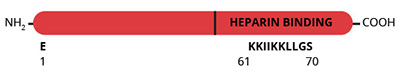
Platelet factor-4 (PF-4) is a low molecular weight, heparin-binding protein which is secreted from agonist-activated platelets (1,2). PF-4 is localized within the platelet a-granule (2) where its concentration ranges from 11.2-12.4 µg per 109 platelets making it, on a molar basis, one of the most abundant proteins in the platelet (1,2). Since the relative concentration of PF-4 in platelets exceeds that of plasma by 280,000-fold (3), PF-4 levels in plasma have been utilized as a measure of platelet activation in vivo. PF-4 is secreted from platelets in complex with a high molecular weight, proteoglycan, carrier protein (4,5). Sedimentation equilibrium experiments have shown that in the absence of its proteoglycan carrier, the molecular weight of PF-4 is 27,000-29,000 (4,5). Subsequent amino acid sequencing of PF-4 revealed a molecular weight of 7,800 (6-8) indicating that native PF-4 is a homotetramer. Functionally, PF-4 neutralizes the anticoagulant activity of heparin in plasma. The heparin binding site within PF-4 is presumably located within the lysine-rich, COOH-terminal region of the molecule (6-8). Since soluble heparin is a therapeutic agent, the physiological significance of the anti-heparin activity of PF-4 is not known. However, by interacting with cell surface expressed heparin-like glycosaminoglycans on endothelial cells, PF-4 may exert its procoagulant effect (9,10). PF-4 binding to cell surface glycosaminoglycans may also be a mechanism through which PF-4 stimulates the release of histamine from basophils (11). The chemotactic activity of PF-4 toward neutrophils and monocytes (12) has also been localized to the COOH-terminal, presumed heparin-binding domain of PF-4 (13). While PF-4 is primarily a secreted protein, PF-4 binding sites on the platelet surface have been identified which may be important for platelet aggregation (14).

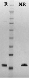
Human PF-4 is prepared from the supernatant of thrombin-activated platelets by heparin-agarose affinity chromatography (15). The purified protein is supplied in 25 mM Hepes pH 7.4, 2 M NaCl and should be stored at -80oC. Purity is assessed by SDS-PAGE analysis and heparin-neutralizing activity is verified by clotting assay.

Sample gel image
LoadHuman Platelet Factor 4, 1 µg per lane
BufferMOPS
StandardSeeBluePlus 2; Myosin (191 kDa), Phosphorylase B (97 kDa), BSA (64 kDa), Glutamic Dehydrogenase (51 kDa), Alcohol Dehydrogenase (39 kDa), Carbonic Anhydrase (28 kDa), Myoglobin Red (19 kDa), Lysozyme (14 kDa)
Localizationplatelet a-granule (2)
Mode of actionneutralizes the anticoagulant activity of heparin. Plasma concentration is used as a marker of platelet activation.
Molecular weight29,000 (4)
Extinction coefficient
E
1 %
1 c m, 280 nm
= 2.6 – calculated based upon amino acid sequence and molecular weight
Isoelectric point7.6 (16)
Structurehomotetramer (monomer, Mr~7800) (5-7)
  1. Rucinski, B., et al., Blood, 53, 47 (1979).
  2. Kaplan, K.L., et al., Blood, 53, 604 (1979).
  3. George, J.N., Blood, 76, 859 (1990).
  4. Barber, A.G., et al., Biochim. Biophys. Acta, 286, 316 (1972).
  5. Moore, S., et al., Biochim. Biophys. Acta, 379, 370 (1975).
  6. Hermodson, M., et al., J. Biol. Chem., 252, 6276 (1977).
  7. Deuel, T.F., et al., Proc. Natl. Acad. Sci. USA, 74, 2256 (1977).
  8. Walz, D.A., et al., Thromb. Res., 11, 833 (1977).
  9. Busch, C., et al., Thromb. Res., 19, 129 (1980).
  10. Rao, A.K., et al., Blood, 61, 1208 (1983).
  11. Brindley, L.L., et al., J. Clin. Invest., 72, 1218 (1983).
  12. Deuel, T.F., et al., Proc. Natl. Acad. Sci. USA, 74, 2256 (1981).
  13. Osterman, D.G., et al., Biochem. Biophys. Res. Commun., 107, 130 (1982).
  14. Capitanio, A.M., et al., Biochim. Biophys. Acta, 839, 161 (1985).
  15. Jordon, R.E., et al., J. Biol. Chem., 257, 400 (1982).
  16. Niewiarowski, S., and Jameson, B., Human Protein Data (A. Haeberli, ed.) © VCH Verlagsges, mbH, Weinheim

No sample publications.

Shopping Cart