Fibronectin

Domain Structure of Fibronectin
The domain structure of the fibronectin monomer is represented. Native fibronectin is composed of two subunits which are linked through two interchain disulfide bonds near the COOH-terminal of each subunit. The transglutaminase (factor XIIIa) crosslinking site is indicated by the arrow. Various macromolecular binding domains, including heparin (HEP), fibrin (FBN), and collagen are shown. The cell adhesion site (CELL) which possesses an RGDS sequence is depicted.

Showing the single result

  • Human Fibronectin

    HCFN-0170

    $196.00

    SKU: HCFN-0170 Category:
    Price:$196.00/2 mg
    Size 2 mg
    Formulation Lyophilized from 20 mM Hepes, 150 mM NaCl, pH 7.4
    Storage 4°C
    Shelf Life 12 months
    Purity >95% by SDS-PAGE
    Activity Determination N/A

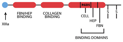
Fibronectin (cold-insoluble globulin) is a high molecular weight, adhesive, glycoprotein found in both plasma (1) and the extracellular matrix (2). Fibronectin is composed of two peptide chains of approximately 275,000 molecular weight which are linked through two interchain disulfide bonds at the COOH-terminal end of the molecule (3). The structure of fibronectin is characterized by three different types of repeating homologous sequence units (4,5). The 45 amino acid type-I repeat constitutes the NH2- and COOH-terminal ends of the protein. The two 60 amino acid type-II segments follow the first nine type-I repeats at the NH2-terminus. The 90 amino acid type-III segments occupy the central region of the fibronectin molecule. Structural differences between plasma and cellular fibronectin (6,7) as well as between the two subunits of plasma fibronectin (8,9) have been identified. These differences likely originate due to transcriptional and posttranscriptional events involving mRNA splicing (10).

The apparent function of fibronectin is to mediate cell attachment by interacting with cell surface receptors and extracellular matrix components (10-12). Fibronectin contains an Arg-Gly-Asp-Ser (RGDS) cell attachment-promoting sequence within one of the type-III homology segments in the middle of the molecule (13). This RGDS site is recognized by specific RGDS-dependent cell receptors (13) which are members of the integrin family of proteins (14). Fibronectin binding to activated platelets (15) is mediated through the RGDS cell adhesion sequence (16). Other binding domains specific for such extracellular macromolecules as heparin, fibrin(ogen), and collagen (10-12) have been identified and may be important in the fibronectin-mediated adhesion of platelets to the extracellular matrix of endothelial cells (17). Fibrin-fibronectin complexes are stabilized by the factor XIIIa-catalyzed covalent cross-linking of fibronectin to the a chain of fibrin (18).

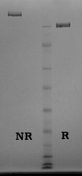
Fibronectin is isolated from human plasma by thermal precipitation, ion exchange, and gelatin-agarose affinity chromatography. The purified protein is supplied as a lyophilizate. Purity is assessed by SDS-PAGE analysis.

Sample gel image
GelNovex 4-12% Bis-Tris
LoadLoad: Human Fibronectin, 1 µg per lane
BufferMOPS
StandardSeeBluePlus 2; Myosin (191 kDa), Phosphorylase B (97 kDa), BSA (64 kDa), Glutamic Dehydrogenase (51 kDa), Alcohol Dehydrogenase (39 kDa), Carbonic Anhydrase (28 kDa), Myoglobin Red (19 kDa), Lysozyme (14 kDa)
Localizationplasma, 0.3 mg/ml (1)
extracellular matrix (2)
Mode of actionmediates cell attachment to extracellular matrix (9-11)
Molecular weight550,000 (3)
Extinction coefficient
E
1 %
1 c m, 280 nm
= 14.0
Isoelectric point~5.0 (19)
Structureheterodimer (3)
Percent carbohydrate4-10%, depending on source (12)
  1. Mosesson, M.W., and Umfleet, R.A., J. Biol. Chem., 245, 5728 (1970).
  2. Stenman, S. and Vaheri, A., J. Exp. Med., 147, 1054 (1978).
  3. Skorstengaard, K., et al., Eur. J. Biochem., 161, 441 (1986).
  4. Kornblihtt, A.R., et al., EMBO J., 4, 1755 (1985).
  5. Odermatt, E., et al., Proc. Natl. Acad. Sci. USA, 82, 6571 (1985).
  6. Hayashi, M. and Yamada, K.M., J. Biol. Chem., 256, 11292 (1981).
  7. Atherton, B.T. and Hynes, R.O., Cell, 25, 133 (1981).
  8. Hayashi, M. and Yamada, K.M., J. Biol. Chem., 258, 3332 (1983).
  9. Sekiguchi, K. and Hakomori, S-I., J. Biol. Chem., 258, 3967 (1983).
  10. Hynes, R.O., Ann. Rev. Cell Biol., 1, 67 (1985).
  11. Mosher, D.F., Ann. Rev. Med., 35, 561 (1984).
  12. Rouslahti, E. Ann. Rev. Biochem., 57, 375 (1988).
  13. Rouslahti, E. and Pierschbacher, M.D., Cell, 44, 517 (1986).
  14. Hynes, R.O., Cell, 48, 549 (1987).
  15. Plow, E.F. and Ginsberg, M.H., J. Biol. Chem., 256, 9477 (1981).
  16. Ginsberg, M., et al., J. Biol. Chem., 260, 3931 (1985).
  17. Houdjik, W.P.M., et al., Arteriosclerosis, 6, 24 (1986).
  18. Mosher, D.F., J. Biol. Chem., 250, 6614 (1975)
  19. Mosher, D.F., Human Protein Data (A. Haeberli, ed.) © VCH Verlagsges, mbH, Weinheim

No sample publications.

Shopping Cart