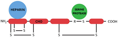
Antithrombin III (ATIII) is a single chain glycoprotein with a molecular weight of 58,000. It is a member of the serpin (serine protease inhibitor) super family and is considered to be the most important inhibitor in the coagulation cascade (1,2). ATIII inhibits a wide spectrum of serine proteases including thrombin, factors IXa, Xa and XIa, kallikrein, plasmin, urokinase, C1-esterase, and trypsin. The mechanism of inhibition involves the formation of a stable 1:1 complex between the active site of the protease and the scissile bond (Arg 385-Ser 386) of ATIII. The active site serine of thrombin has been shown to form a covalent intermediate with the P1 amino acid (Arg 385) of ATIII. The rate of inhibition of serine proteases by ATIII is increased to varying degrees by heparin. In the case of being a thrombin inhibitor or factor Xa inhibitor, the interaction with ATIII is enhanced 3 orders of magnitude in the presence of heparin. The interaction between ATIII and heparin involves a unique sequence of sulfated and non-sulfated monosaccharide units on heparin, and critical lysine residues on ATIII. The binding of ATIII to heparinoid structures on vascular endothelium has been demonstrated and shown to enhance the inhibition of factors IXa, Xa, and thrombin.
ATIII may also function in the complement cascade. The binding of ATIII to fluid phase complement attack-complexes in sera has been demonstrated. In addition, the S protein of complement (an inhibitor of the membrane attack-complex) interferes with the ATIII/thrombin interaction.
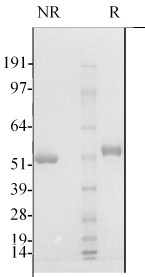
ATIII is prepared from fresh frozen plasma by heparin-agarose affinity chromatography (3). The purified protein is supplied in 50% (vol/vol) glycerol/H2O and should be stored at -20°C. Purity is determined by SDS-PAGE analysis.