Human Coagulation Factor XII

Domain Structure of Factor XII
The organization of the factor XII domain structure based on sequence homology is represented, where: EGF=epidermal growth factor, Type I and Type II=domains homologous to those found in fibronectin, solid arrows=kallikrein cleavage sites that form a- and β-factor XIIa, dashed arrows=cleavages leading to intermediate forms of factor XIIa.

Showing the single result

  • Human Factor XII

    HCXII-0155

    $69.00$530.00

    SKU: HCXII-0155 Category:
    Price:$69.00/100 µg, $530.00/1mg
    Size 100 µg, 1mg
    Formulation 50% glycerol/4mM Sodium Acetate, 0.15M NaCl, pH 5.3 (v/v)
    Storage -20°C
    Shelf Life 12 months
    Purity >95% by SDS-PAGE
    Activity Determination Clotting assay

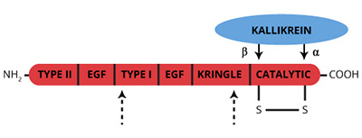
Factor XII (XII) (Hageman Factor) is a single chain (Mr=78,000) glycoprotein zymogen that circulates in plasma at a concentration of 40 µg/ml (1-5). Reciprical activation of XII to the active serine protease factor XIIa (XIIa) by kallikrein is central to initiation of the intrinsic coagulation pathway. Surface bound α-XIIa in turn activates factor XI to XIa. Secondary cleavage of α-XIIa by kallikrein yields β-XIIa, which catalyzes solution phase activation of kallikrein, factor VII and the classical complement cascade.

The ability of a variety of negatively charged substances, both physiological and nonphysiological to promote XII activation and, thus, initiation of the intrinsic pathway has led to the psuedonym “contact activation”. Binding to anionic surfaces induces a conformational change, making the XII zymogen more susceptible to cleavage by a variety of proteases (6,7). It is unlikely that binding to negatively charged surfaces alone is sufficient to activate XII, since highly purified preparations of XII and plasma deficient in prekallikrein and high molecular weight kininogen do not undergo this “autocatalysis” (8-11).

A single cleavage by kallikrein at R353-Val354 of XII yields α-XIIa, a 2 chain protease (Mr=80,000) held together by disulfide bonds. The COOH-terminal light chain (Mr=28,000) contains the catalytic triad (His-40, Asp-89, Ser-191), while the NH2-terminal heavy chain (Mr=52,000) conatins the anionic surface binding portion of the molecule. A secondary cleavage of α-XIIa by kallikrein outside the disulfide bond yields β-XIIa (XIIf, BHFa, HFf, hageman factor fragments) (Mr=28,000), which no longer binds anionic surfaces (12). β-XIIa can activate prekallikrein, but has little procoagulant activity (13,14). Several other minor intermediate forms of XIIa are indicated in the figure above.

Inhibitors of XIIa include C1-INH, α2-antiplasmin, α2-macroglobulin and antithrombin III. At physiological concentrations, the relative effectiveness of these inhibitors is 91 : 4.5 : 3 : 1.5, respectively (10, 16-19). The ratio of C1-INH to XII has been implicated in the “cold activation” of factor VII and the conversion of prorenin to renin on storage of plasma (20,21).

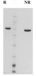
Human factor XII is prepared from fresh frozen plasma by immunoaffinity chromatography and supplied in 50% glycerol/4mM Sodium Acetate, 0.15M NaCl, pH 5.3 for storage at -20oC.

Sample gel image
GelNovex 4-12% Bis-Tris
LoadHuman Factor XII, 1 µg per lane
BufferMOPS
StandardSeeBluePlus 2; Myosin (191 kDa), Phosphorylase B (97 kDa), BSA (64 kDa), Glutamic Dehydrogenase (51 kDa), Alcohol Dehydrogenase (39 kDa), Carbonic Anhydrase (28 kDa), Myoglobin Red (19 kDa), Lysozyme (14 kDa)
Special NotesHeavy chain is a doublet due to the presence of up to 50% beta form. The conversion of alpha-Xa to beta-Xa occurs by autocleavage of alpha-Xa by alpha-Xa resulting in the loss of a COOH-terminal peptide.
LocalizationPlasma
Plasma concentration40 µg/ml (3)
Mode of actionZymogen; precursor to the serine protease factor XIIa; activated by kallikrein/HMWK/anionic surface complex to intitiate the intrinsic pathway
Molecular weight80,000 (2)
Extinction coefficient
E
1 %
1 c m, 280 nm
= 14.0
Isoelectric point6.8
Structuresingle chain (mr=80,000), organized into 6 domains based on sequence homology (5).
Percent carbohydrate17%
  1. Schmaier, A.H., et al., in Hemostasis and Thrombosis, ed. R.W. Colman, J. Hirsh, V.J. Marder and E.W. Salzman, pp 18-38, J.B. Lippincott Company, Philadelphia, 1987.
  2. Griffin, J.H. and Cochrane, C.G., Methods Enzymol., 45, 56-65 (1976).
  3. Saito, H., et al., J Lab Clin., 88, 506 (1976).
  4. McMullen, B.A. and Fujikawa, N., J. Biol. Chem., 260, 5328 (1985).
  5. Davie, E.W., in Hemostasis and Thrombosis, ed. R.W. Colman, J. Hirsh, V.J. Marder and E.W. Salzman, pp 242-267, J.B. Lippincott Company, Philadelphia, 1987.
  6. Griffin, J.H., Proc. Natl. Acad. Sci. USA, 75, 1998 (1978).
  7. McMillin, L.R., et al., J. Clin. Invest., 54, 1312 (1974).
  8. Revak, S.D., et al., J. Clin. Invest., 59, 1167 (1977).
  9. Fujikawa, K., et al., Biochemistry, 16, 4182 (1977).
  10. Bonno, N., et al., in New Comprehensive Biochemistry, ed. R.F.A. Zwaal and H.C.Hemker, pp. 103-128, Elsevier, Amsterdam, 1986.
  11. Claeys, H. and Collen, D. (1978) Eur. J. Biochem., 87, 69 (1978).
  12. Revak, S.D. and Cochrane, C.G., J. Clin. Invest., 57, 852 (1976).
  13. Cochrane, C.G., et al., J. Exp. Med., 138, 1564 (1973).
  14. Revak, S.D., et al., J. Exp. Med., 147, 719 (1978).
  15. Ghebrehiwet, B., et al., J. Clin. Invest., 71, 1458 (1983).
  16. DeAgostini, A., et al., J. Clin. Invest., 73, 1542 (1984).
  17. Rathoff, O.D., et al., J. Exp. Med., 129, 315 (1969).
  18. Stead, N., et al., J. Biol Chem., 251, 6481 (1976).
  19. Pixley, R.A., et al., J. Biol Chem., 260, 1723 (1985).
  20. Gjonnass, H., Thromb. Diath. Haemorrh., 28, 182 (1972).
  21. Radcliffe, R., et al., Blood, 59, 611 (1977).
  1. Choudhri, T., et al., J Exp Med. 1999 July 1; 190(1): 91–100. (Activation of factor IX)

This publication list is not all encompassing, and is only meant to provide limited examples of how Prolytix products are used. We encourage you to search the literature for other examples pertinent to your experimentation, and to contact us with any technical questions.

Shopping Cart