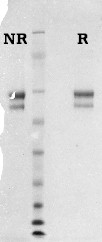
Vitronectin is a plasma glycoprotein that circulates in the blood at a concentration range of 200 to 400 mg/ml constituting 0.2 to 0.5% of total plasma proteins (1,2). It is detected as a mixture of both 75 kDa and 65 kDa forms (3). The 65 kDa form is a product of proteolytic processing of the former which appears to be endogenously present. Vitronectin is involved in a number of physiological processes that include blood coagulation, fibrinolysis, complement cascade, and cell adhesion. Vitronectin (Serum spreading factor) is one of the two major cell-adhesive glycoproteins. It is a major ligand for the vitronectin receptor (αV/β3) or αV/β5 integrin on adhesive cells (4). Other functions include heparin-binding; collagen-binding; osteonectin-binding; complement lysis inhibitor and “scavenger” protein for macromolecular products of the complement and haemostasis cascade systems (5). It also functions as a substrate for trans glutaminase (Factor XIIIa) crosslinking, cAMP-dependent protein kinase, and tyrosyl protein sulfotransferase (4,5). Vitronectin has implications for thrombosis due to its heparin binding properties. It has been postulated that vitronectin prevents the acceleration of clot formation inhibition by acting as a heparin scavenger ultimately protecting thrombin and factor Xa from heparin-dependant inactivation by antithrombin III or heparin cofactor II (6,7). Vitronectin is also thought to play an important role in fibrinolysis by its ability to bind active PAI-1 (4). Extracellular binding of vitronectin to plasminogen activator inhibitor-1 (PAI-1) and plasminogen, stabilize the inhibitor and thus affect tissue plasminogen activator-mediated plasmin formation (4).
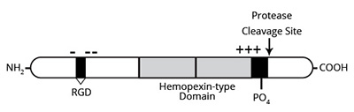
The vitronectin domain structure starting at the NH4-terminus consists of a somatomedin-B region (residues 1 to 44), a connecting segment containing the major cell attachment RGD sequence (residues 45 to 47) and a highly acidic region (residues 53 to 64), followed by hemopexin-like repeats (homology to hemopexin which is a heme-binding plasma protein), a glycosaminoglycan-binding site which is represented by a 40-amino acid stretch rich in basic residues, a protease-sensitive region susceptible to high concentrations of thrombin, and a COOH-terminal protein kinase A-dependent phosphorylation site, which is adjacent to the protease cleavage site (4,5).
Human vitronectin is prepared from fresh frozen plasma similar to the procedure described by Yatohgo and coworkers (8). Purified vitronectin is supplied in 50 mM sodium phosphate, 150 mM NaCl, pH 7.4 and should be stored at -80°C. Purity is assessed by SDS-PAGE analysis.