Beta-Thromboglobulin

Domain Structure of Beta Thromboglobulin
The domain structure of the β-thromboglobulin monomer is represented. The β-thromboglobulin monomer is an 8,800 molecular weight peptide which is derived via NH2-terminal proteolysis of a precursor molecule LAPF-4 (low affinity platelet factor-4). Although the COOH-terminal domain of β-thromboglobulin is characterized by a clustering of basic lysine residues, the affinity of b-thromboglobulin for heparin is significantly weaker than that of platelet factor-4. In its native state, β-thromboglobulin is a homotetramer consisting of four, identical, noncovalently-

Showing the single result

  • Human Beta-Thromboglobulin

    HBTG-0210

    Size 1 mg, 100 µg
    Formulation 25 mM Hepes, 150 mM NaCl, pH 7.4
    Storage -80°C
    Shelf Life 12 months
    Purity >95% by SDS-PAGE
    Activity Determination N/A

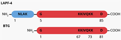
β-thromboglobulin (b-TG), is a low molecular weight, heparin-binding, platelet-derived protein (1). It is similar to platelet factor-4 (PF-4) in that it is localized within the platelet alpha-granule at levels reported to range from 8.1-24.2 µg per 109 platelets (2,3). The relative concentration of β-TG in platelets exceeds that of plasma by 260,000-fold (4) making β-TG a convenient marker of platelet activation. Structurally, β-TG is analogous to PF-4 in that, in its native state, β-TG is a tetramer (1) consisting of four identical 8800 molecular weight peptide chains (5). In contrast to PF-4, β-TG exhibits a lower affinity for heparin and also exists as a larger molecular weight species known as “low affinity PF-4” (LAPF-4) (2). β-TG is derived from the proteolytic removal of four NH2-terminal amino acid residues from a LAPF-4 (6,7). Immunological screening of partially fractionated supernatant from activated platelets revealed a highly basic form of β-TG distinct from LAPF-4 (7). This basic β-TG species, termed platelet basic protein (PBP), was subsequently isolated (8) and later concluded from immunological, peptide sequencing, and proteolytic processing studies to be a higher molecular weight precursor form of both LAPF-4 and β-TG (9,10).


The physiological function of β-TG is not known. While early studies suggested that the precursor forms of β-TG were mitogenic for mouse fibroblasts (8,11), it was later concluded that this activity was due to growth factor contamination (10). β-TG has also been reported to inhibit prostacyclin-I2 production by endothelial cells (12), however, the relevance of this effect has been called into question (13,14). The chemotactic activity of platelet alpha-granule proteins for human fibroblasts has been attributed to both PF-4 and β-TG (15).

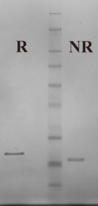
Human β-TG is prepared from the supernatant of activated platelets by heparin-agarose affinity chromatography and gel filtration (1,2). The purified protein is supplied in 25 mM Hepes, 150 mM NaCl pH 7.4 and should be stored at -80°C. Purity is assessed by SDS-PAGE analysis.

Sample gel image
GelNovex 4-12% Bis-Tris
LoadLoad: Human Beta Thromboglobulin, 1 µg per lane
BufferMES
StandardSeeBluePlus 2; Myosin (188 kDa), Phosphorylase B (98 kDa), BSA (62 kDa), Glutamic Dehydrogenase (49 kDa), Alcohol Dehydrogenase (38 kDa), Carbonic Anhydrase (28 kDa), Myoglobin Red (17 kDa), Lysozyme (14 kDa), Aprotinin (6 kDa), Insulin, B chain (3 kDa).
Localizationplatelet alpha-granule (3)
Plasma Concentration100-200 µg/ml
Mode of actionheparin-binding protein: Plasma concentration used as a marker of platelet activation
Molecular weight35,800 (1)
Extinction coefficient
E
1 %
1 c m, 280 nm
= 2.6 (calculated based upon amino acid sequence and molecular weight)
Structurehomotetramer (monomer, Mr~8800) (5)
  1. Moore, S., et al., Biochim. Biophys. Acta, 379, 360 (1975).
  2. Rucinski, B., et al., Blood, 53, 47 (1979).
  3. Kaplan, K.L., et al., Blood, 53, 604 (1979).
  4. George, J.N., Blood, 76, 859 (1990).
  5. Begg, S., et al., Biochemistry, 17, 1739 (1978).
  6. Holt, J.C. and Niewiarowski, S., Biochim. Biophys. Acta, 632, 284 (1980).
  7. Niewiarowski, S., et al., Blood, 55, 453 (1980).
  8. Paul, D., et al., Proc. Natl. Acad. Sci. USA, 77, 5914 (1980).
  9. Varma, K.G., et al., Biochim. Biophys. Acta, 701, 7 (1982).
  10. Holt, J.C., et al., Biochemistry, 25, 1988 (1986).
  11. Niewiarowski, S. and Paul, D., in Platelets in Biology and Pathology, Vol. 2, pp. 91-106, (Gordon, J.L. ed.) Elsevier/North-Holland Biomedical Press (1981).
  12. Hope, W., et al., Nature, 282, 210 (1979).
  13. Ager, A. and Gordon, J.L., Thromb. Res., 24, 95 (1981).
  14. Poggi, A. et al., Proc. Soc. Exp. Biol. Med., 172, 543 (1983).
  15. Senior, R.M., et al., J. Cell. Biol., 96, 382 (1983).

No sample publications.

Shopping Cart