Human Coagulation Factor VIIa

Participation of Factor VIIa in the Extrinsic Factor Xase Complex
The components of the extrinsic factor Xase enzyme complex are illustrated. Factor VIIa and its cofactor tissue factor (TF) assemble on a negatively charged membrane surface in a calcium dependent manner to form an enzyme complex which proteolytically converts factor X to factor Xa. The complex is referred to as the extrinsic factor Xase complex, because the tissue factor component is presumably derived from a source “extrinsic” to the plasma environment, which is accessible following vascular injury.

Showing the single result

  • Human Factor VIIa

    HCVIIA-0031

    Size 1 mg, 20 µg
    Formulation 50% glycerol/water (v/v)
    Storage -20°C
    Shelf Life 12 months
    Purity >95% by SDS-PAGE
    Activity Determination Factor VII clotting assay

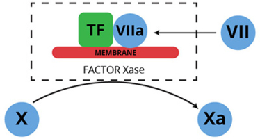
Human factor VII is a single chain plasma glycoprotein, and is a zymogen in its native form (1-4). Proteolytic activation of factor VII, yields the enzyme factor VIIa, which when bound to the integral membrane protein tissue factor, forms an enzyme complex that proteolytically converts factor X to factor Xa. This enzyme complex is best known as the extrinsic factor Xase (pronounced ten-ase) complex, since by virtue of its tissue factor component, it is composed of protein normally extrinsic to the plasma environment.

The conversion of factor VII to factor VIIa is catalyzed by a number of proteases including thrombin, factor IXa, factor Xa, factor XIa, and factor XIIa. Rapid activation also takes place when factor VII is combined with tissue factor in the presence of calcium (5-9). This latter event is consistent with autocatalysis, and is likely initiated by a small amount of pre-existing factor VIIa. The activation reaction results in cleavage of the peptide bond between arginine 152 and isoleucine 153.

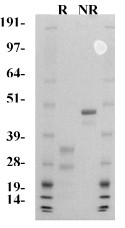
The resulting factor VIIa consists of an NH2-derived light chain (Mr=20,000), and a COOH-terminal derived heavy chain (Mr= 30,000), which remain associated through a single disulfide bond (cys 135 – cys 262). The light chain contains the membrane binding “Gla domain”, while the heavy chain contains the catalytic domain.

Unlike other serine proteases, factor VIIa alone is not readily inhibited by the antithrombin III/heparin complex. However, in the presence of tissue factor, antithrombin III/heparin exhibits significant inhibition of factor VIIa (6).

Human factor VIIa is prepared from affinity purified factor VII, and purified by ion exchange chromatography. The purified protein is supplied in 50% (vol/vol) glycerol/H2O and should be stored at -20°C. The purity is determined by SDS-PAGE analysis, and activity is measured in a factor VII clotting assay.

Sample gel image
GelNovex 4-12% Bis-Tris
LoadHuman Factor VIIa, 1 µg per lane
BufferMOPS
StandardSeeBluePlus 2; Myosin (191 kDa), Phosphorylase B (97 kDa), BSA (64 kDa), Glutamic Dehydrogenase (51 kDa), Alcohol Dehydrogenase (39 kDa), Carbonic Anhydrase (28 kDa), Myoglobin Red (19 kDa), Lysozyme (14 kDa)
LocalizationPlasma
Mode of actionEnzyme component of the extrinsic factor X activating complex; also activates factor IX, thus by-passing the contact activation system.
Molecular weight50,000 (human) (2)
Extinction coefficient
E
1 %
1 c m, 280 nm
= 13.9 (2) – (inferred from the zymogen, factor VII)
Specific Activityapproximately 16,000 units/mg (determined from single stage clotting assay)
Structure2 subunits; NH2-terminal derived light chain (Mr=20,000), COOH-terminal derived heavy chain (Mr=30,000), NH2-terminal gla-domain, two EGF domains
Percent carbohydrate13% (10) – (based upon analysis of bovine factor VII)
Post-translational modificationsone β-hydroxyaspartate (11), ten gla residues (12)
  1. Broze, G.J. and Majerus, P.W., Methods Enzymol., 80, 228 (1981).
  2. Bajaj, S.P., et al., J. Biol. Chem., 256, 253 (1981).
  3. Williams, E.B., et al., J. Biol. Chem., 264, 7536 (1989).
  4. Kisiel, W. and McMullen, B.A., Thrombosis Res., 22, 375 (1981).
  5. Radcliffe, R. and Nemerson, Y., J. Biol. Chem., 250, 388 (1975).
  6. Lawson, J.H.,et al., J. Biol. Chem., 268, 767 (1993).
  7. Kisiel, W., et al., Biochemistry, 16, 4189 (1977).
  8. Radcliffe, R., et al., Blood, 50, 611 (1977).
  9. Seligsohn, U., et al., J. Clin. Invest., 64, 1056 (1979).
  10. Kisiel, W., et al., Biochemistry, 14, 4928 (1975).
  11. McMullen, B.A., et al., BBRC, 115, 8 (1983).
  12. Discipio, R.G., et al., Biochemistry, 16, 698 (1977).
  1. Gopinath, S., et al., Biochem. J. (2007) 405, 351–357. (Tissue Factor-VIIa)
  2. Maroney, S., et al., Blood. 2007 March 1; 109(5): 1931–1937. (Tissue Factor-VIIa)

This publication list is not all encompassing, and is only meant to provide limited examples of how Prolytix products are used. We encourage you to search the literature for other examples pertinent to your experimentation, and to contact us with any technical questions.

Shopping Cart