Domain Structure of Protein S
The domain structure of protein S is represented, where: GLA = region containing γ-carboxyglutamic acid residues, EGF = region containing sequences homologous to human epidermal growth factor, TSR = thrombin sensitive region, ? = region of unknown function which replaces the catalytic triad found in vitamin K-dependent serine proteases.

Showing the single result

  • Human Protein S

    HCPS-0090

    PLEASE INQUIRE

    SKU: HCPS-0090 Category:
    Size 1 mg, 100 µg
    Formulation 50% glycerol/water (v/v)
    Storage -20°C
    Shelf Life 12 months
    Purity >95% by SDS-PAGE
    Activity Determination N/A

Protein S is a single chain vitamin K-dependent protein which is thought to function in both the coagulation and complement cascades (1,2). Approximately 60% of protein S circulating in plasma is complexed to C4b binding protein (C4BP). It has been suggested that g-carboxyglutamic acid (gla) dependent binding of protein S to negatively charged phospholipids may function to concentrate C4BP at cell surfaces following injury.

In the coagulation system, protein S functions as an anticoagulant cofactor protein. Activated protein C (APC) forms a 1:1 stoichiometric complex with protein S in the presence of Ca2+ and phospholipid vesicles (Kd=6×10⁻⁹M) (3). In the presence of protein S, a moderate increase (3-10 fold) in the rate of factor Va and factor VIIIa inactivation by APC is observed in plasma and on the surface of unstimulated platelets. Protein S bound to C4BP does not possess APC cofactor activity. Recently, an additional binding protein which enhances the activity of protein S has been described (4). Proteolytic inactivation of protein S by thrombin has been proposed as a regulatory mechanism in this system. A single cleavage by thrombin abolishes protein S cofactor activity by removing an NH2-terminal peptide (Mr=8000) which contains the gla domain.

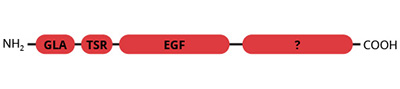
The domain structure of protein S is similar to that of the other vitamin K-dependant coagulation factors with the exception that protein S does not possess the catalytic triad. Protein S is a single chain protein containing 10 gla residues in the NH2-terminal domain and 4 epidermal growth factor (EGF) domains.

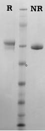
Human protein S is isolated from fresh frozen plasma by a combination of conventional methods (9) and immunoaffinity chromatography as described by Jenny et al. (5). Purified protein S is supplied in 50% (vol/vol) glycerol/H2O and should be stored at -20°C. Purity is determined by SDS-PAGE analysis.

Sample gel image
LoadHuman Protein S, 1 µg per lane
BufferMOPS
StandardSeeBluePlus 2; Myosin (191 kDa), Phosphorylase B (97 kDa), BSA (64 kDa), Glutamic Dehydrogenase (51 kDa), Alcohol Dehydrogenase (39 kDa), Carbonic Anhydrase (28 kDa), Myoglobin Red (19 kDa), Lysozyme (14 kDa)
LocalizationPlasma, free and complexed to C4BP
Plasma concentration10 µg/ml (free) (6)
Mode of actionCofactor for activated protein C
Molecular weight69,000 (7)
Extinction coefficient
E
1 %
1 c m, 280 nm
= 9.5 (7)
Isoelectric point5.0-5.5 (7)
Structuresingle chain, NH2-terminal gla domain, four EGF domains
Percent carbohydrate7% (7)
Post-translational modificationsone β-hydroxyaspartate (8) ten gla residues (7), three β-hydroxyasparagine (8)
  1. Walker, F.J., Semin. Thromb. Hemostas., 10, 131 (1984).
  2. Dahlback, B., et al., Semin. Thromb. Hemostas., 10, 139 (1984).
  3. Walker, F.J., J. Biol. Chem., 256, 11128 (1981).
  4. Walker, F.J., J. Biol. Chem., 261, 10941 (1986).
  5. Jenny, R.J., et al., Prep. Biochem., 16, 227 (1986).
  6. Dahlback, B., Biochem. J., 209, 837 (1983).
  7. Discipio, R.G., et al., Biochemistry, 18, 899 (1979).
  8. Stenflo, J., et al., Proc. Natl. Acad. Sci. USA, 84, 368 (1987).
  9. Bajaj, S.P., et al., Prep. Biochem., 13, 191 (1983).

No sample publications.

Shopping Cart