Bovine Lactadherin

The Structure of Lactadherin
Lactadherin (MFG-E8) binds, Ca++-independently, to phosphatidyl-L-serine via the C2 domain and binds the avb3 and avb5 integrins via the second EGF domain. The one letter amino acid codes refer to identified amino acids that participate in binding. For the C2 domain the capital letters refer to water-exposed hydrophobic residues.

Showing all 2 results

  • Bovine Lactadherin

    BLAC-1200

    $181.00

    SKU: BLAC-1200 Category:
    Price:$181.00/50 ug
    Size 50 ug
    Formulation 70 mM Sodium Phosphate, pH 7.0
    Storage -80°C
    Shelf Life 12 months
    Purity >95% by SDS-PAGE
    Activity Determination N/A
  • Bovine Lactadherin - FITC Labeled

    BLAC-FITC

    $786.00

    SKU: BLAC-FITC Category:
    Price:$786.00/1 ml
    Size 1 ml
    Formulation TBS, 1% BSA, 0.02% azide, pH 7.4
    Storage 4°C
    Shelf Life 12 months
    Purity >95% by SDS-PAGE
    Activity Determination N/A

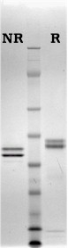
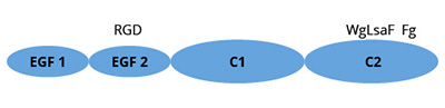
Lactadherin is a widely distributed glycoprotein (~ 50 kDa), which was originally characterized due to its association with milk fat/lipid globule membranes. Synonymous names are PAS-6/7, bovine-associated mucoprotein, BA-46, P47, and MFG-E8. Structural hallmarks of lactadherin are the presence of two epidermal growth factor (EGF) homology domains (with an RGD peptide motif in the second EGF domain), and two C domains sharing homology with the discoidin family of lectin domains including the phospholipid-binding domains of blood clotting factors V and VIII. Lactadherin shows preferential binding to phosphatidylserine (L-form) in a calcium independent manner, and binds more specifically than Annexin V.

Purified lactadherin functions as an anticoagulant by blocking phosphatidylserine-containing membrane sites for blood coagulation proteins (10). Fluoresence-labeled lactadherin functions as a sensitive probe for exposed phosphatidylserine on nucleated cells and on stimulated platelets (8, 9) . Lactadherin will bind to membranes that have phosphatidylserine content below the threshold for annexin V binding.

Lactadherin is purified from un-pasteurized bovine milk (11).

Illustrated Applications

K562 cells stained with lactadherin and annexin V

HL60 cellsstained with lactadherin and annexin V

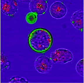
Above: K562 cells (top) and HL60 cells (bottom) co-stained with both FITC-conjugated lactadherin (green) and Alexa-647 conjugated annexin V (red) early in apoptosis. The annexin is internalized in granules and is not detectably staining the cells.  Reference: Shi, J., Y. Shi, L. N. Waehrens, J. T. Rasmussen, C. W. Heegaard and G. E. Gilbert (2006). “Lactadherin detects early phosphatidylserine exposure on immortalized leukemia cells undergoing programmed cell death.” Cytometry A 69(12): 1193-201.  Copyright 2006.  John Wiley & Sons, Inc.  Reprinted with permission of John Wiley & Sons, Inc.

HeLa cells stained with Lactadherin after treatment with staurosporine 2 hour

HeLa cells stained with Lactadherin after treatment with staurosporine 3 hour

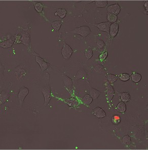
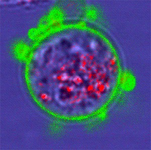
Above: HeLa cells stained with FITC-conjugated lactadherin 2 hours (top) and 3 hours (bottom) after treatment with staurosporine. Early in apoptosis the cells have small vesicles and long, thin appendages that stain avidly for lactadherin. Reference: Waehrens LN, Heeghaard, CW, Gilbert GE, Rasmussen JT. Bovine Lactadherin as a Calcium-independent Imaging Agent of Phosphatidylserine Expressed on the Surface of Apoptotic HeLa Cells 2009 J. Histochem. Cytochem. (ePub June 2009).


Mesenteric-venous-thrombosis


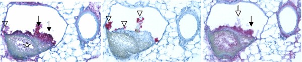
Above: Phosphatidylserine exposure in mouse mesenteric venous thrombosis.  Mice were given 1 µg each of lactadherin and annexin V by tail vein immediately prior to externalization of the mesentery. The mesentary was exposed to ferric chloride and then the animals were perfused with saline/paraformaldehyde. Serial sections of the mesentary were stained with anti-fibrinogen/fibrin (left), anti-platelet (middle), and anti-lactadherin antibodies (right) developed with the alkaline phosphatase Vector Red substrate. A layer of fibrinogen/fibrin (left, closed arrows) overlaid a mural hemorrhage (open star). Platelets (middle) were scattered along the luminal surface of the thrombus (open triangles) as well as upon fibrinogen/fibrin strands extending into the lumen. Lactadherin staining (right) was strongest along the raised endothelium surface (closed arrow), including adherent platelets close to the wall. Platelets on fibrin strands did not stain detectably (open arrow). (Shi J, Pipe SW, Rasmussen JT, Heegaard CW, Gilbert GE. Lactadherin blocks thrombosis and hemostasis in vivo: correlation with platelet phosphatidylserine exposure. J Thromb Haemost. Jul 2008;6(7):1167-1174).

Guidelines for using BLAC-FITC:

FITC-conjugated bovine lactadherin (BLAC-FITC) is supplied as a 100X stock solution (1.6 micromolar) in a buffer of 20mM Tris, 150mM NaCl, pH 7.4 containing 1% (w/v) bovine serum albumin and 0.02% sodium azide.  Assuming that most labeling reaction volumes will be approximately 0.5ml, a 1 ml vial of the 100X stock material will be sufficient for 200 labeling reactions.  Additionally, as this product is fluorescently labeled it should be protected from light.

For a typical cell staining experiment, apoptotic cells are collected by centrifugation and  resuspended in a physiologic buffer such as TBS (20mM Tris, 150mM NaCl, pH 7.4), HBS (20mM Hepes, 150mM NaCl, pH 7.4) or PBS (4.3mM Na2HPO4, 1.47mM KH2PO4, 137mM NaCl, 2.7 mM KCL, pH 7.4) to a final cell count of approximately 1 x 10⁶ cells/ml.  Adherent cells may be harvested by trypsinization, but should be washed at least once in either media or buffer prior to making the final suspension in buffer.  Stock 100X FITC-conjugated lactadherin is added to the cell suspension at the rate of 5 microliters for every 0.5ml of the cell suspension.  At this point, optional staining with propidium iodide (PI) may be initiated by adding PI to a final concentration of  0.5 to 1 µg/ml.  Incubate the reaction mixture at room temperature (protected from light) for a period of 5 to 10 minutes.

Labeled cells may be analyzed by a variety of methods including flow cytometry and fluorescence microscopy.   Fluorescence detection may be monitored using the following detector settings:

BLAC-FITC:      Ex = 488 nm; Em = 530 nm

PI:                    Ex = 488 nm; Em = 640 nm

Sample gel image
GelNovex 4-12% Bis-Tris
LoadBovine Lactadherin, 1 µg per lane
BufferMOPS
StandardSeeBluePlus 2; Myosin (191 kDa), Phosphorylase B (97 kDa), BSA (64 kDa), Glutamic Dehydrogenase (51 kDa), Alcohol Dehydrogenase (39 kDa), Carbonic Anhydrase (28 kDa), Myoglobin Red (19 kDa), Lysozyme (14 kDa)
NamesLactadherin, MFG-E8, PAS-6/7, SED1, p47
LocalizationMilk (1) , also secreted by stimulated macrophages (2) , vascular smooth muscle (3) , endothelial cells (4)
Plasma concentrationunknown in normal adults, measurable in women with metastatic breast cancer (5) , measurable in newborn calves after milk meal (1)
Mode of actionPhysiologic function as opsonin or bridge ligand (6, 7) . The lectin-like domains engage phosphatidyl-L-serine, the second EGF domain engages avb3 and/or avb5 integrin leading to phagocytosis of apoptotic cells.
Molecular weight52 kDa and 47 kDa (glycosylation variants, PAS-6 and PAS-7 respectively)
Extinction coefficient
E
1 %
1 c m, 280 nm
= 16.5 (calculated)
Structuresingle chain with two EGF domains and two C domains. The mouse isoform contains a central proline rich region and the human molecule contains a single EGF domain
Percent carbohydrate6%, 13% for glycosylation variants
Post-translational modificationsglycosylation
  1. Butler, J.E., Pringnitz, D.J., Martens, C.L., and Crouch, N. 1980. Bovine-associated mucoprotein: I. Distribution among adult and fetal bovine tissues and body fluids. Differentiation 17:31-40.
  2. Hanayama, R., Tanaka, M., Miwa, K., Shinohara, A., Iwamatsu, A., and Nagata, S. 2002. Identification of a factor that links apoptotic cells to phagocytes. Nature 417:182-187.
  3. Haggqvist, B., Naslund, J., Sletten, K., Westermark, G.T., Mucchiano, G., Tjernberg, L.O., Nordstedt, C., Engstrom, U., and Westermark, P. 1999. Medin: an integral fragment of aortic smooth muscle cell-produced lactadherin forms the most common human amyloid. Proc Natl Acad Sci U S A 96:8669-8674.
  4. Silvestre, J.S., Thery, C., Hamard, G., Boddaert, J., Aguilar, B., Delcayre, A., Houbron, C., Tamarat, R., Blanc-Brude, O., Heeneman, S., et al. 2005. Lactadherin promotes VEGF-dependent neovascularization. Nat Med 11:499-506.
  5. Ceriani, R.L., Sasaki, M., Sussman, H., Wara, W.M., and Blank, E.W. 1982. Circulating human mammary epithelial antigens in breast cancer. Proc Natl Acad Sci U S A 79:5420-5424.
  6. Hanayama, R., Tanaka, M., Miyasaka, K., Aozasa, K., Koike, M., Uchiyama, Y., and Nagata, S. 2004. Autoimmune disease and impaired uptake of apoptotic cells in MFG-E8-deficient mice. Science 304:1147-1150.
  7. Hanayama, R., and Nagata, S. 2005. Impaired involution of mammary glands in the absence of milk fat globule EGF factor 8. Proc Natl Acad Sci U S A 102:16886-16891.
  8. Shi, J., Shi, Y., Waehrens, L.N., Rasmussen, J.T., Heegaard, C.W., and Gilbert, G.E. 2006. Lactadherin detects early phosphatidylserine exposure on immortalized leukemia cells undergoing programmed cell death. Cytometry A 69:1193-1201.
  9. Dasgupta, S.K., Guchhait, P., and Thiagarajan, P. 2006. Lactadherin binding and phosphatidylserine expression on cell surface-comparison with annexin A5. Transl Res 148:19-25.
  10. Shi, J., and Gilbert, G.E. 2003. Lactadherin inhibits enzyme complexes of blood coagulation by competing for phospholipid binding sites. Blood 101:2628-2636.
  11. Hvarregaard et al., 1996. Eur. J. Biochem. 240.

Other references of interest:

1.         Akakura S, Singh S, Spataro M, Akakura R, Kim JI, Albert ML, et al. The opsonin MFG-E8 is a ligand for the alphavbeta5 integrin and triggers DOCK180-dependent Rac1 activation for the phagocytosis of apoptotic cells. Exp Cell Res. 2004 Jan 15;292(2):403-16. PubMed PMID: 14697347.

2.         Baghdadi M, Chiba S, Yamashina T, Yoshiyama H, Jinushi M. MFG-E8 regulates the immunogenic potential of dendritic cells primed with necrotic cell-mediated inflammatory signals. PLoS One. 2012;7(6):e39607. PubMed PMID: 22761839. Pubmed Central PMCID: 3382463.

3.         Cui T, Miksa M, Wu R, Komura H, Zhou M, Dong W, et al. MFG-E8 Attenuates Acute Lung Injury in Mice after Intestinal Ischemia and Reperfusion. Am J Respir Crit Care Med. 2009 Nov 5. PubMed PMID: 19892861.

4.         Hanayama R, Tanaka M, Miyasaka K, Aozasa K, Koike M, Uchiyama Y, et al. Autoimmune disease and impaired uptake of apoptotic cells in MFG-E8-deficient mice. Science. 2004 May 21;304(5674):1147-50. PubMed PMID: 15155946.

5.         Jinushi M, Nakazaki Y, Dougan M, Carrasco DR, Mihm M, Dranoff G. MFG-E8-mediated uptake of apoptotic cells by APCs links the pro- and antiinflammatory activities of GM-CSF. J Clin Invest. 2007 Jul;117(7):1902-13. PubMed PMID: 17557120.

6.         Mobarrez, F. et al. Microparticles in the blood of patients with systemic lupus erythematosus (SLE): phenotypic characterization and clinical associations. Sci. Rep. 6, 36025; doi: 10.1038/srep36025 (2016).

7.         Rothmeier, A. et al. Caspase-1–mediated pathway promotes generation of thromboinflammatory microparticles. J Clin Invest. 2015 Apr 1; 125(4): 1471–1484.

Shopping Cart