Domain Structure of Protein C
The domain structure of protein C is represented, where: GLA = region containing γ-carboxyglutamic acid residues, EGF = region containing sequences homologous to human epidermal growth factor, AP = activation peptide released upon conversion of the zymogen to the active serine protease, CATALYTIC DOMAIN = region containing the serine protease catalytic triad. The arrow indicates the site which is proteolytically cleaved by thrombin during activation of the zymogen.

Showing all 2 results

  • Bovine Protein C

    BCPC-1070

    $106.00$980.00

    SKU: BCPC-1070 Categories: ,
    Price:$106.00/100 µg, $980.00/1mg
    Size 100 µg, 1mg
    Formulation 50% glycerol/water (v/v)
    Storage -20°C
    Shelf Life 12 months
    Purity >95% by SDS-PAGE
    Activity Determination < 0.5% aPC activity by chromogenic assay
  • Human Protein C

    HCPC-0070

    $191.00$1,498.00

    SKU: HCPC-0070 Categories: ,
    Price:$191.00/100 µg, $1,498.00/1mg
    Size 100 µg, 1mg
    Formulation 50% glycerol/water (v/v)
    Storage -20°C
    Shelf Life 12 months
    Purity >95% by SDS-PAGE
    Activity Determination < 0.5% aPC activity by chromogenic assay

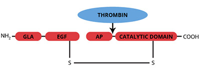
The vitamin K-dependent zymogen, protein C, is synthesized in the liver as a single chain polypeptide and is subsequently converted to a disulfide linked heterodimer, by removal of a dipeptide (Lys-146 and Arg-147) from the precursor molecule (1,2). Trace quantities of the single chain form have been observed in plasma. The light chain, which is responsible for the calcium dependent binding of protein C to phospholipid vesicles, contains 11 γ-carboxyglutamic acid (gla) residues, 1 b-hydroxyaspartic acid residue, and 2 epidermal growth factor (EGF) homology domains. The serine protease catalytic triad is located in the heavy chain. Human protein C is susceptible to proteolytic cleavage of a peptide (Mr=3000) from the COOH-terminal end of the heavy chain, yielding an altered form referred to as β-protein C. No functional distinction between α- and β-protein C has been observed. A single cleavage at Arg-12 (Arg-14 in bovine) of the heavy chain of human protein C converts the zymogen into the serine protease, activated protein C. This cleavage is catalyzed by a complex between α-thrombin and the endothelial cell surface protein thrombomodulin. In contrast to the other vitamin K dependent coagulation factors, activated protein C functions as an anticoagulant by catalyzing the proteolytic inactivation of factors Va and VIIIa. APC also contributes to the fibrinolytic response by complex formation with plasminogen activator inhibitors.

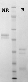
Bovine protein C is prepared from fresh citrated bovine plasma by a modification of the Walker procedure (3), as described by Haley et al. (4). Human protein C is prepared from fresh frozen citrated human plasma using a combination of immunoaffinity chromatography (5), and conventional techniques (4,9). Protein C is provided in 50% (vol/vol) glycerol/H2O and should be stored at -20oC. Purity is determined by SDS-PAGE analysis and activity is measured using a chromogenic substrate based assay.

Sample gel image
LoadHuman Protein C, 1 µg per lane
BufferMOPS
StandardSeeBluePlus 2; Myosin (191 kDa), Phosphorylase B (97 kDa), BSA (64 kDa), Glutamic Dehydrogenase (51 kDa), Alcohol Dehydrogenase (39 kDa), Carbonic Anhydrase (28 kDa), Myoglobin Red (19 kDa), Lysozyme (14 kDa)
LocalizationPlasma
Plasma concentration4-5 µg/ml (human) (6)
5-10 µg/ml (bovine) (2)
Mode of actionZymogen; precursor to the serine protease activated protein C (APC)
Molecular weight62,000 (human) (7)
58,000 (bovine) (7)
Extinction coefficient
E
1 %
1 c m, 280 nm
= 14.5 (human) (7)
  = 13.7 (bovine) (7)
Isoelectric point4.4-4.8 (human) (8)
4.2-4.5 (bovine) (8)
Structuretwo chains, Mr=41,000 and 21,000, disulfide linked, NH2-terminal gla domain two EGF domains
Percent carbohydrate23 % (human) (7)
14 % (bovine) (7)
Post-translational modificationseleven gla residues (bovine), nine gla residues (human), one β-hydroxyaspartate
  1. 1. Esmon, C.T., Progress in Thromb. and Hemosts., 10, 25 (1984).
  2. Stenflo, J., Semin. in Thromb. and Hemostas., 10, 109 (1984).
  3. Walker, F.J., et al., Biochim. Biophys. Acta, 571, 333 (1979).
  4. Haley, P.E., et al., J. Biol. Chem., 264, 16303 (1989).
  5. Jenny, R.J., et al., Prep. Biochem., 16, 227 (1986).
  6. Griffen, J.H., et al., Blood, 60, 261 (1982).
  7. Kisiel, W., et al., Methods Enzymol., 80, 320 (1981).
  8. Discipio, R.G., et al., Biochemistry, 18, 899 (1979).
  9. Bajaj, S.P., et al., Prep. Biochem., 11, 397 (1981).
  1. Hwang, K., et. al., Arthritis Rheum. 2003 June ; 48(6): 1622–1630. (used as ELISA capture)

This publication list is not all encompassing, and is only meant to provide limited examples of how Prolytix products are used. We encourage you to search the literature for other examples pertinent to your experimentation, and to contact us with any technical questions.

Shopping Cart