Activated Protein C

Inactivation of Factor Va by Activated Protein C (APC)
Proteolytic inactivation of factor Va by APC is represented, where: PS=protein S, PCPS=phospholipid vesicles (or cellular surface) and Ca++=calcium ions. The inactivation of factor Va to form factor Vai results from proteolysis of the factor Va heavy chain (94K) at three specific sites by APC (solid arrows) (1,2). The location of these cleavage sites in the factor Va heavy chain are as follows: (human: R306, R506, & R679) and (bovine: R306, R505, & R662). Complete inactivation of the cofactor molecule requires cleavage at the Arginine-306 position. Cleavage at Arginine-306 by activated protein C occurs only in the presence of membrane, and requires prior cleavage of the heavy chain at Arginine-505. Proteolysis of the factor Va light chain by APC occurs only in the bovine molecule and is not required for inactivation (dashed arrow).

Showing all 2 results

  • Bovine Activated Protein C

    BCAPC-1080

    Price:$1,440.00/1mg, $82.00/50 µg
    Size 1mg, 50 µg
    Formulation 50% (vol/vol) Glycerol/H2O
    Storage -20°C
    Shelf Life 12 months
    Purity >95% by SDS-PAGE
    Activity Determination Chromogenic Assay
  • Bovine Activated Protein C - DEGR (active site blocked)

    BCAPC-DEGR

    Price:$1,700.00/1mg, $94.00/50 µg
    Size 1mg, 50 µg
    Formulation 20 mM Hepes, 150 mM NaCl, pH 7.4
    Storage -80°C
    Shelf Life 12 months
    Purity >95% by SDS-PAGE
    Activity Determination Chromogenic Assay

Activated protein C (APC) is an anticoagulant serine protease derived from the two chain, vitamin K-dependent zymogen, protein C (3-7). A complex between alpha-thrombin and thrombomodulin catalyzes a single cleavage at Arg-12 (Arg-14 in bovine) in the heavy chain of protein C, to generate activated Protein C. Several non-physiologically relevant proteases such as RVV-X activator, trypsin, and PROTAC are also capable of activating protein C.

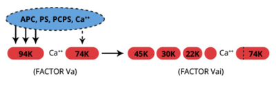
APC functions as an anticoagulant which catalyzes the proteolytic inactivation of the cofactors, factors Va and VIIIa, leading to inhibition of the prothrombinase and factor Xase complexes. The inactivation of factors Va and VIIIa is both Ca2+ and phospholipid dependent. The vitamin K dependent cofactor, protein S, moderately increases this rate of inactivation by forming a 1:1 complex with APC (Kd=6×10⁻⁹M) (8).

Several factors attenuate the anticoagulant activity of APC. Factor Xa protects factor Va from proteolysis by APC by competing for a similar binding site on factor Va. Thrombin has also been proposed as a regulator of APC by proteolytic inactivation of protein S. In addition, APC is regulated by a circulating heparin-dependent protein C inhibitor (PAI-3), a circulating heparin-independent protein C inhibitor, a platelet-derived protein C inhibitor, and PAI-1. The complexes formed between APC and both types of PAI have been reported to account for increased fibrinolysis observed upon infusion of APC or the generation of APC in vivo.

In addition to our standard APC preparation, an active site-blocked form containing Dansyl-EGR-chloromethlyketone is also available.

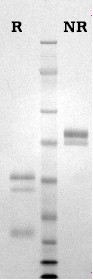
Activated protein C is prepared from purified protein C by activation with thrombin followed by ion exchange chromatography (4). APC is supplied in 50% (vol/vol) glycerol/H2O and should be stored at -20oC. Purity is determined by SDS-PAGE analysis and activity is measured using a chromogenic substrate assay. All production lots of APC are also tested for their ability to prolong the aPTT of normal human plasma, as required for the APC resistance assay (10,11). The results of this test are provided for each lot, as an aPTT (+/- APC) ratio (10nM APC).


Sample gel image
GelNovex 4-12% Bis-Tris
LoadHuman aPC, 1 µg per lane
BufferMOPS
StandardSeeBluePlus 2; Myosin (191 kDa), Phosphorylase B (97 kDa), BSA (64 kDa), Glutamic Dehydrogenase (51 kDa), Alcohol Dehydrogenase (39 kDa), Carbonic Anhydrase (28 kDa), Myoglobin Red (19 kDa), Lysozyme (14 kDa)
LocalizationPlasma
Mode of actionAnticoagulant, inactivates factors Va and VIIIa
Molecular weight56,200 (human) (5)
52,650 (bovine) (5)
Extinction coefficient
E
1 %
1 c m, 280 nm
= 14.5 (human) (9)
  = 13.7 (bovine) (9)
Isoelectric Point4.4-4.8 (human) (9)
4.2-4.5 (bovine) (9)
Structuretwo chains, Mr=35,000 and 21,000, disulfide linked, NH2-terminal gla domain two EGF domains
Percent carbohydrate23 % (human) (5)
14 % (bovine) (5)
Post-translation modificationseleven gla residues (bovine), nine gla residues (human), one β-hydroxyaspartate
  1. Kalafatis, M., and Mann, K.G., J. Biol. Chem., 268, 27246 (1993).
  2. Kalafatis, M., et al., J. Biol. Chem., 269, 31869 (1994).
  3. Esmon, C.T., Progress in Thromb. and Hemostas., 10, 25 (1984).
  4. Esmon, C.T., J. Biol. Chem., 264, 4743 (1989).
  5. Kisiel, W., et al., Methods Enzymol., 80, 320 (1981).
  6. Stenflo, J., Semin. in Thromb. and Hemostas., 10, 109 (1984).
  7. Marlar, R.A., Semin. in Thromb. and Hemostas., 11, 387 (1985).
  8. Walker, F.J., et al., J. Biol. Chem., 256, 11128 (1981).
  9. Discipio, R.G., et al., Biochemistry, 18, 899 (1979).
  10. Dahlback, B., and Hildebrand, B., Proc. Natl. Acad. Sci. USA, 91, 1396 (1994).
  11. Svensson, P.J., and Dahlback, B., New Engl. J. Med., 330, 517 (1994).
  1. Hwang, K., Arthritis Rheum. 2003 June ; 48(6): 1622–1630. (used as capture in ELISA)
  2. Raife, T., et al., J Clin Invest. 1994 April; 93(4): 1846–1851. (used as standard in aPC generation)
  3. Schuepbach, R., et al., Blood. 2008 March 1; 111(5): 2667–2673. (PAR1 cleavage)
  4. Adams, T., Hockin, M., Mann, K., Everse, S., Proc Natl Acad Sci U S A. 2004 June 15; 101(24): 8918–8923. (bovine aPC used to inactivate bovine Va)
  5. Gale, A. et al., J. Biol. Chem. (2008) VOL. 283, NO. 24, pp. 16355–16362. (Inactivation of VIIIa)

This publication list is not all encompassing, and is only meant to provide limited examples of how Prolytix products are used. We encourage you to search the literature for other examples pertinent to your experimentation, and to contact us with any technical questions.

Shopping Cart