Human Coagulation Factor VII

Domain Structure of Human Factor VII
The domain structure of human factor VII is represented, where: GLA = region containing γ-carboxyglutamic acid residues, EGF = region containing sequences homologous to human epidermal growth factor, CATALYTIC DOMAIN = region containing the serine protease catalytic triad. The site at which factor Xa cleaves factor VII to form factor VIIa is indicated with an arrow.

Showing the single result

  • Human Factor VII

    HCVII-0030

    Price:$7,569.00/1mg, $191.00/20 µg
    Size 1mg, 20 µg
    Formulation 50% glycerol/water (v/v)
    Storage -20°C
    Shelf Life 12 months
    Purity >95% by SDS-PAGE
    Activity Determination Clotting assay

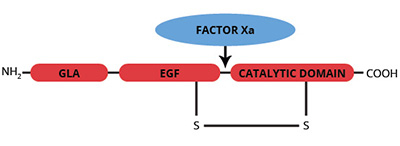
Human factor VII is a single chain, vitamin K-dependent, plasma glycoprotein which is synthesized in the liver (1-3). Prior to secretion into the blood, post translational modification by a vitamin K-dependent carboxylase produces ten-carboxyglutamic acid (gla) residues located in the NH2-terminal portion of the molecule, which facilitate cell membrane binding. Factor VII is proteolytically activated to the serine protease, factor VIIa, during coagulation. Factor VII can be activated by thrombin, factor IXa, factor Xa or factor XIIa. The activation results in cleavage of the single chain molecule on the COOH-terminal side of arginine-152, to produce an NH2-terminal derived light chain (Mr=20,000) and a COOH-terminal derived heavy chain (Mr=30,000) which remain covalently associated by a single disulfide bond. The light chain region contains the gla domain, as well as two growth factor domains which are homologous to human epidermal growth factor (EGF). A single β-hydroxyaspartic acid identified in factor VII is also located in the light chain region. The heavy chain region of factor VIIa contains the catalytic domain. Factor VIIa and the cofactor, tissue factor, may combine on negatively charged cell surfaces in a calcium dependent manner to form the extrinsic factor Xase enzyme complex. This enzyme complex catalyzes the conversion of both factor IX to factor IXa and factor X to factor Xa. The cDNA for factor VII has been isolated and the nucleotide sequence determined (4). Factor VII shares extensive sequence homology with other serine proteases including factor IX, factor X and protein C.

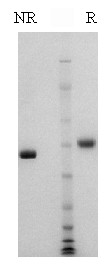
Human factor VII is purified using a combination of conventional techniques (2) and immunoaffinity chromatography (5). The purified protein is supplied in 50% (vol/vol) glycerol/H2O and should be stored at -20oC. Purity is determined by SDS-PAGE analysis and activity is measured in a factor VII clotting assay.

Sample gel image
GelNovex 4-12% Bis-Tris
LoadHuman Factor VII, 1 µg per lane
BufferMOPS
StandardSeeBluePlus 2; Myosin (191 kDa), Phosphorylase B (97 kDa), BSA (64 kDa), Glutamic Dehydrogenase (51 kDa), Alcohol Dehydrogenase (39 kDa), Carbonic Anhydrase (28 kDa), Myoglobin Red (19 kDa), Lysozyme (14 kDa)
LocalizationPlasma
Plasma concentration0.5 µg/ml (2) – (based upon activity measurements)
Mode of actionZymogen; precursor to the serine protease factor VIIa
Molecular weight50,000 (2)
Extinction coefficient
E
1 %
1 c m, 280 nm
= 13.9
Isoelectric point4.8-5.1 (6) – (based upon analysis of bovine factor VII)
Structuresingle chain, NH2-terminal gla-domain, two EGF domains
Percent carbohydrate13% (7) – (based upon analysis of bovine factor VII)
Post-translational modificationsone β-hydroxyaspartate (8), ten gla residues (9)
  1. Davie, E.W., et al., Adv. Enzymol., 48, 277 (1979).
  2. Bajaj, S.P., et al., J. Biol. Chem., 256, 253 (1981).
  3. Broze, G.J., et al., J. Biol. Chem., 255, 1242 (1980).
  4. Hagen, F.S., et al., Proc. Natl. Acad. Sci., USA, 83, 2412 (1986).
  5. Jenny, R.J., et al., Prep. Biochem., 16, 227 (1986).
  6. Discipio, R.G., et al., Biochemistry, 18, 899 (1979).
  7. Kisiel, W., et al., Biochemistry, 14, 4928 (1975).
  8. McMullen, B.A., et al., BBRC, 115, 8 (1983).
  9. Discipio, R.G., et al., Biochemistry, 16, 698 (1977).

No sample publications.

Shopping Cart