Beta-2-Glycoprotein 1

Domain Structure of Beta-2-Glycoprotein 1
The five contiguous Sushi domains ( I-V ) of β2I are shown. The 5 carbohydrate side chains at N73, N143, N164, N174, and N224 are represented (CHO).

Showing all 2 results

  • Beta2-Glycoprotein I

    B2GI-0001

    $106.00$920.00

    Price:$920.00/1 mg, $106.00/100 µg
    Size 1 mg, 100 µg
  • Human Beta-2-Glycoprotein 1

    B2G1-0001

    $103.00

    SKU: B2G1-0001 Category:
    Price:$103.00/100 µg
    Size 100 µg
    Formulation Lyophilized from 0.2 M Glycine, 150 mM NaCl, pH 7.4
    Storage 4°C
    Shelf Life 12 months
    Purity >95% by SDS-PAGE
    Activity Determination Plasma equivalency units

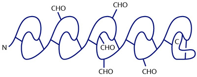
β2-Glycoprotein I (β2I, Apolipoprotein H) is a highly glycosylated single chain protein (Mr=54,200) which is synthesized in the liver and circulates in plasma at a concentration of 100-200 µg/ml (1-4). Approximately 40% of the plasma β2I is associated with lipoproteins, which led to the designation apolipoprotein H (5). The 326 amino acid protein contains five repeating mutually homologous domains consisting of approximately 60 amino acids which are disulfide bonded to form Short Consensus Repeats (SCR) or Sushi domains. The third, C-terminal sushi domain contains three of the five N-linked carbohydrate chains, at positions N143, N164, N174. The other two sites are at N73 and N234 in domains II and IV, respectively. There are 38 basic amino acid residues distributed unevenly among the five domains; eight in domain I, five in domain II, five in domain III, five in domain IV and fifteen in domain V. The direct binding of domains I and V has been demonstrated and likely results from folding of the molecule in a manner that expresses a net cationic surface charge (5).

Although the physiological role of β2I is still being studied, the ability to bind to anionic surfaces has been the focus of intense investigation. β2I has been shown to bind to anionic vesicles (3), platelets (6), DNA (7), mitochondria (8) and heparin (9). It has been suggested that the binding of β2I to negatively charged surfaces can inhibit the contact activation pathway in blood coagulation. The binding to activated platelets is reported to inhibit platelet associated prothrombinase and adenylate cyclase activities. Studies on physiologic roles for binding to DNA, mitochondria, heparanoids and phospholipids have focused on the area of autoimmune disorders. In particular, the complexes between β2I and cardiolipin have been implicated in the anti-phospholipid related disorders LAC and SLE (10-13).

The human β2-Glycoprotein I is purified from fresh frozen human plasma, using a combination of ion exchange and affinity chromatography. In addition it is purified over an anti-human factor XI column to remove any potential contaminents of XI/XIa. It is greater than 95% pure by SDS PAGE. The human β2-Glycoprotein I is lyophilized from a glycine-NaCl buffer and should be stored at 4°C.

Sample gel image
GelNovex 4-12% Bis-Tris
LoadHuman Beta-2 Glycoprotein 1, 1 µg per lane
BufferMOPS
StandardSeeBluePlus 2; Myosin (191 kDa), Phosphorylase B (97 kDa), BSA (64 kDa), Glutamic Dehydrogenase (51 kDa), Alcohol Dehydrogenase (39 kDa), Carbonic Anhydrase (28 kDa), Myoglobin Red (19 kDa), Lysozyme (14 kDa)
LocalizationPlasma
Plasma Concentration100-200 µg/ml
Mode of actionCationic residues on the surface of sushi domains I and V bind to anionic phospholipids. Inhibition of contact activation, platelet membrane dependant prothrombinase and adenylate cyclase activities. Complexes with DNA, mitochondria, and cardiolipin augments antigenicity in autoimmune disorders.
Molecular weight54,200 (SDS PAGE), 48,000 (Sed. Equil)
Extinction coefficient
E
1 %
1 c m, 280 nm
= 10.0
Structure326 amino acid single chain protein with 5 contiguous Short Consensus Repeats on sushi domains.
Percent carbohydrate25%
Post-translational modifications5 N-linked glycosylation sites, 1 in domain II (N73), 3 in domain III (N143, N164, N174) and 1 in domain IV (N224).
  1. Lozier, J., et.al., Proc.Natl.Acad. Sci.,USA, 81, 2640-3644 (1984).
  2. Kato, H. and Enjyoi, K., Biochem., 30, 11687-11694 (1997).
  3. Wurm, H., Int.J.Biochem., 16, 511-515 (1984).
  4. Bendixen, E., et.al., Biochem., 31, 3611-3617 (1992).
  5. Steinkasserer, A., et.al., Biochem.J., 277, 387-391 (1991).
  6. Nimpf, J. et.al., Biochim.Biophys.Acta, 884, 142-149 (1986).
  7. Kroll, J. et.al., Biochim.Biophys.Acta, 434, 490-501 (1986).
  8. Schousboe, I., Biochim.Biophys.Acta, 679, 396-408 (1979).
  9. Polz, E., et.al., Int.J.Biochim., 11, 265-273 (1076).
  10. McNeil, H.P. et.al., Proc.Natl.Acad.USA, 87, 4120-4124 (1990).
  11. Galli, M. et.al., Lancet, I, 1544-1547 (1990).
  12. Matsuuna, E., et.al., Lancet, II, 177-178 (1990).
  13. Pengo, V. et.al., Thrombosis and Haemostasis, 73, 29-34 (1995).
  1. Shi, T., et al., Proc Natl Acad Sci U S A. 2004 March 16; 101(11): 3939–3944.
  2. Reed, J.H. et al., Arthritis & Rheumatism, Vol. 60, No. 3, March 2009, pp 860–869 (DOI 10.1002/art.24361) – used in with apoptosis cells)

This publication list is not all encompassing, and is only meant to provide limited examples of how Prolytix products are used. We encourage you to search the literature for other examples pertinent to your experimentation, and to contact us with any technical questions.

Shopping Cart